
Decellularized bile duct scaffold in tissue engineering: is it suitable for biliary tract surgery?
Massaro M.S., Sevcik J., Palek R., Panova E., Sarcevic S., Matsviayonak I., Bolek L., Liska V., Moulisova V.
Bile duct reconstruction after injury or resection is a common procedure of hepatopancreatobiliary surgery. The current method is the reconstruction of the bile duct with a piece of intestine. This technique, despite reaching a good compromise for the patient, might result in the return of intestine content towards the liver, causing inflamation and/or obstruction. Research to find suitable bile duct substitutes for anatomical reconstruction has been carried out over the last 100 years without reaching an optimal result. Our idea is to use porcine bile ducts as a scaffold for reconstrution. The native bile duct is decellularized, meaning that all cells are removed to leave a non-immunogenic scaffold. A scheme of the experiment is shown in Figure 1. To test if the idea is promising, scaffolds are implanted in vivo as bile ducts in a porcine model and the animals are monitored for 28 days. The results show the absence of an immune reaction at the implantation site and a spontaneous migration of cells in the decellularized scaffold. Furthermore, to verify the compatibility of this porcine scaffold with human cells, a bioreactor, shown in Figure 2, was developed to mimic in vitro the in vivo conditions and seed mesenchymal stem cells on the lumen of the scaffold. The bioreactor provided the mechanical cues for the cells
to completely cover the inner lumen and orient in the direction of the flow.
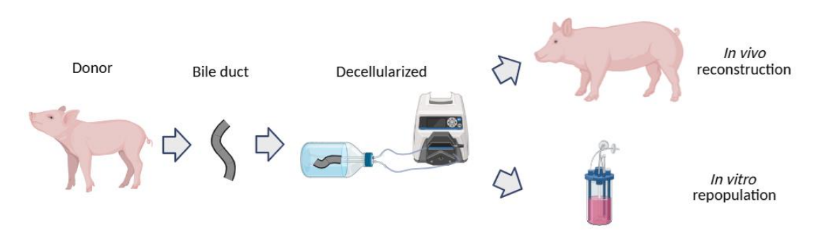
Figure 1 – Scheme of the research: The bile duct is explanted from the donor, decellularized with detergents in perfusion and then the graft is rather implanted in vivo or used for cells seeding in vitro.
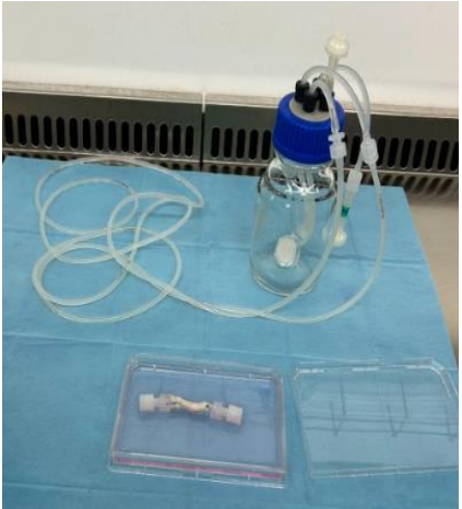
Figure 2 – In vitro repopulation: The cells are seeded in the lumen of the bile duct and then a in-house developed bioreactor is used to mimic the in vivo flow to stimulate cell spreading and proliferation.